Data Overview:
This 133-subject dataset (1.47TB) was acquired for and analyzed in Kronemer et al. 2022. The primary aim of this study was to investigate the cortical and subcortical networks for visual conscious perception. The dataset consists of two adult healthy participants groups: (1) 3T MRI anatomical and BOLD images (TR = 1000ms; voxel size = 2x2x2mm) and (2) 256-channel scalp EEG (sampling rate = 1000Hz). Concurrent pupillometry and eye-tracking (EyeLink 1000 Plus; sampling rate = 1000Hz) is available for most participants. The dataset includes adult epilepsy patient participants with concurrent depth EEG and 20-channel scalp EEG recordings. All recordings were made concurrently with a custom visual perception task.
Citation:
Kronemer, S.I., Aksen, M., Ding, J.Z. et al. Human visual consciousness involves large scale cortical and subcortical networks independent of task report and eye movement activity. Nat Commun 13, 7342 (2022). https://doi.org/10.1038/s41467-022-35117-4
Dataset Summary:
The dataset contains 4 different types of data acquisition experiments:- No Report Paradigm: Center Relevant and Quadrant Relevant MRI + EEG Sessions
- Report-Only Paradigm MRI: Quadrant Relevant MRI-only Session
- Report-Only Paradigm EEG: Quadrant Relevant EEG-only Session
- Report-Only Paradigm depthEEG: Quadrant Relevant depthEEG-only Session
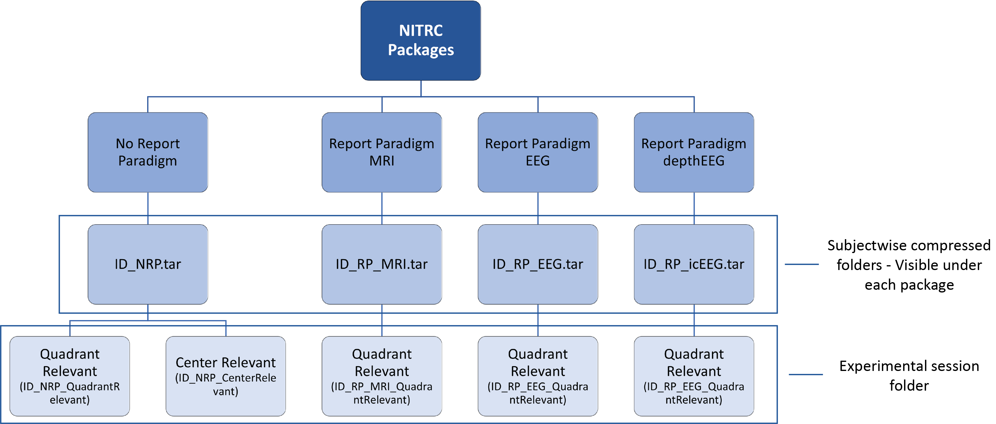
Dataset on NITRC:
The data for each subject for the 4 different types of data acquisition experiments are organized in .tar files and packaged in 4 different packages:- No Report Paradigm
- Report Paradigm MRI
- Report Paradigm EEG
- Report Paradigm depthEEG
The subject name for each experimental session is denoted as {subject ID}_{Paradigm abbreviation}_{data acquisition modality (only for Report Paradigm)}.
 b)
b) 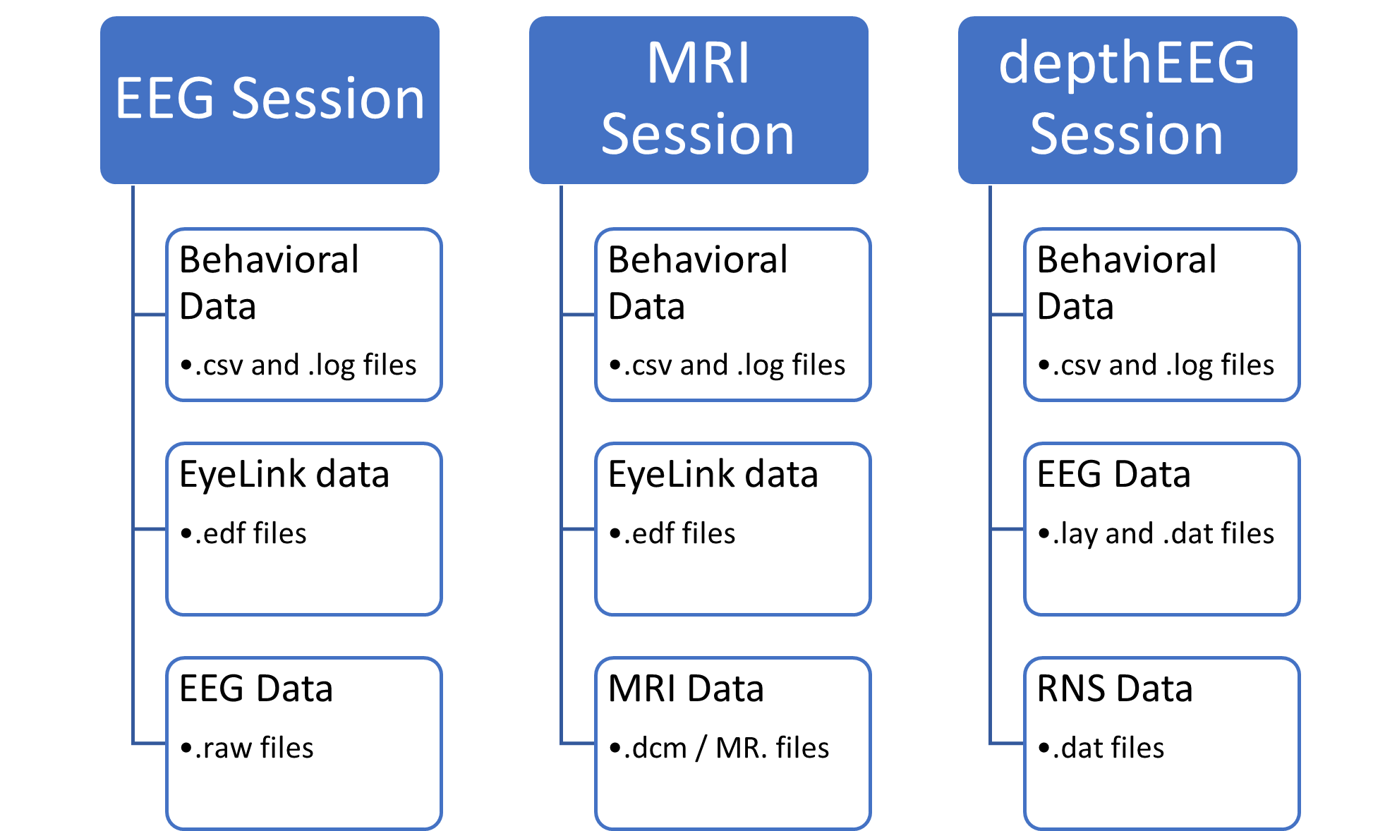
Notes: The MRI MPRAGE images for some MRI sessions were defaced as DICOM files and some were defaced after converting to NIFTI. The defaced DICOMS are in the MRI_Data folder in MRI_seesion. But, the defaced NIFTI images are in MRI_data/Defaced_MPRAGE_NIFTI folder. Also, for some subjects, one of the the experimental session did not take place and thus those data are not available. Additionally, for some subjects, one or more data acquisition sessions did not occur and those sessions were left out in the dataset.
Data availability:
Subject-wise downloads page: Click this button to access the data on NITRC's page: Click this button to access the analysis codes: Click this button to access the Extended Data Slides:

This work is licensed under a Creative Commons Attribution 4.0 International License.